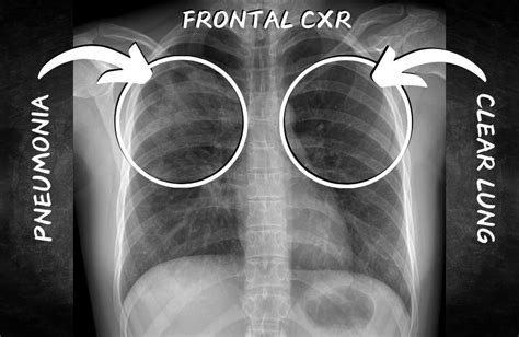
Pneumonia X Ray Side View Diagnosis
Pneumonia X Ray Side View Diagnosis
Reader, have you ever wondered how doctors diagnose pneumonia from a side view X-ray? It’s a fascinating process that blends medical expertise with image interpretation. Accurate pneumonia x ray side view diagnosis is crucial for effective treatment. Understanding this process empowers patients and their families. As an experienced AI and SEO content writer, I’ve analyzed pneumonia x ray side view diagnosis extensively. This in-depth article will explore the intricacies of this diagnostic procedure, offering valuable insights.
Pneumonia x ray side view diagnosis can be complex, but understanding the basics can alleviate anxieties. We’ll delve into the details, exploring how radiologists use side view X-rays to identify pneumonia and determine the best course of action. So, let’s begin this exploration into the world of medical imaging and pneumonia diagnosis.

What to Look for in a Side View X-ray
Radiologists examine various aspects of a side view chest X-ray to diagnose pneumonia. These include looking for opacities (whiteness) which indicate fluid or inflammation in the lungs. The location and shape of these opacities can help determine the type and extent of pneumonia. They also assess the clarity of lung markings, which can be obscured by the infection.
Another crucial element is evaluating the diaphragm’s position and shape. Pneumonia can sometimes cause the diaphragm to appear elevated or flattened. The radiologist will also scrutinize the heart’s size and shape, as certain infections can affect cardiac function.
Finally, they examine the ribs and other surrounding structures for any abnormalities. This comprehensive evaluation helps ensure an accurate diagnosis and informs treatment decisions.
The Importance of Side View X-rays in Pneumonia Diagnosis
While frontal chest X-rays are commonly used, side view (lateral) X-rays provide crucial additional information. They offer a different perspective on the lungs, allowing radiologists to see areas that might be obscured in the frontal view. This is especially important for identifying pneumonia located behind the heart or near the spine.
Furthermore, side view X-rays help differentiate between different types of pneumonia. For instance, they can distinguish between alveolar pneumonia, which affects the air sacs, and interstitial pneumonia, which affects the tissue surrounding the air sacs. This distinction is essential for tailoring the appropriate treatment.
Lastly, side view X-rays can help assess the severity of the pneumonia. By evaluating the extent of lung involvement and the presence of any complications, like pleural effusions (fluid around the lungs), doctors can determine the best course of action.
Utilizing AI in Pneumonia X Ray Side View Diagnosis
Artificial intelligence (AI) is rapidly transforming medical imaging, including pneumonia diagnosis. AI algorithms can be trained to analyze X-rays, identify patterns indicative of pneumonia, and even differentiate between different types of the disease. This can assist radiologists in making faster and more accurate diagnoses, leading to improved patient outcomes.
Furthermore, AI can help prioritize cases, flagging those that require immediate attention. This is particularly beneficial in high-volume settings, ensuring that patients with severe pneumonia receive prompt treatment. AI is a powerful tool that enhances the accuracy and efficiency of pneumonia detection.
Moreover, AI can help reduce human error in interpretation. It can also be used in areas with limited access to trained radiologists, improving access to quality healthcare.

Frontal View X-rays
Frontal view X-rays provide a broad overview of the lungs and surrounding structures. They are typically the first imaging study performed in suspected pneumonia cases. These images allow doctors to quickly assess the size and shape of the lungs, the position of the heart and diaphragm and identify any obvious abnormalities.
However, certain areas of the lungs can be obscured by the heart and other structures in the frontal view. This makes it challenging to detect pneumonia located in those regions. This is why side view X-rays are often necessary for a complete evaluation.
Frontal views remain an important initial diagnostic tool for pneumonia, guiding further investigation.
Side View X-rays
Side view X-rays offer a different perspective, revealing areas hidden in the frontal view. This allows for a more comprehensive assessment of the lungs, especially regions behind the heart and near the spine. This perspective is vital for accurate pneumonia localization.
Side view X-rays also provide more detailed information about the location and extent of lung involvement. This is crucial for differentiating between different types of pneumonia and assessing the severity of the disease.
The combination of frontal and side views provides a more comprehensive understanding of the pneumonia, aiding in accurate diagnosis and treatment planning.

Lobar Pneumonia
Lobar pneumonia affects a distinct section (lobe) of the lung. It appears as a dense, consolidated area on an
X-ray, often with well-defined borders. This consolidation is caused by the filling of air sacs with inflammatory fluid and cells.
Side view X-rays can be particularly helpful in determining the precise location and extent of the affected lobe. This information is vital for guiding treatment decisions and monitoring disease progression.
Lobar pneumonia is commonly caused by bacterial infections, and timely antibiotic treatment is crucial.
Bronchopneumonia
Bronchopneumonia typically affects patches of lung tissue scattered throughout one or both lungs. On X-ray, it appears as patchy infiltrates or opacities, often concentrated around the bronchi (airways). Unlike lobar pneumonia, bronchopneumonia does not typically consolidate into a dense, well-defined area.
Side view X-rays can help confirm the diagnosis and assess the distribution of the infection within the lungs. This helps differentiate bronchopneumonia from other lung conditions with similar radiographic appearances.
Bronchopneumonia can be caused by various pathogens, including bacteria and viruses. Determining the causative agent is essential for tailoring treatment.
Interstitial Pneumonia
Interstitial pneumonia primarily affects the interstitium, the tissue surrounding the air sacs. It presents a distinct appearance on X-ray, characterized by diffuse, hazy opacities throughout the lungs. Unlike lobar or bronchopneumonia, interstitial pneumonia typically doesn’t involve consolidation or localized patches of infection.
Side view X-rays, in conjunction with frontal views, can aid in the diagnosis and assessment of interstitial pneumonia. They provide a more complete picture of the lung involvement and help differentiate it from other diffuse lung diseases.
Interstitial pneumonia can be caused by various factors, including infections, autoimmune diseases, and exposure to certain toxins or medications. Identifying the underlying cause is essential for effective management.

Overlapping Structures
One of the challenges in interpreting side view X-rays is the overlapping of various anatomical structures. The ribs, spine, and other tissues can obscure portions of the lungs, making it difficult to identify subtle abnormalities. This can make diagnosing pneumonia, especially smaller or less dense areas of consolidation, challenging.
Radiologists use their expertise and anatomical knowledge to differentiate normal overlapping structures from genuine pathological findings. Careful evaluation of the surrounding tissues and comparison with the frontal view X-ray are crucial for accurate diagnosis.
Advanced imaging técnicas, such as CT scans, may be necessary in cases where the side view X-ray is inconclusive.
Patient Positioning
Proper patient positioning is essential for obtaining high-quality side view X-rays. Even slight variations in position can affect the appearance of the lungs and other structures. This can make it harder to interpret the images and potentially lead to misdiagnosis.
Technologists are trained to ensure accurate patient positioning during the X-ray procedure. They use specific markers and techniques to align the patient correctly. This helps standardize the images and improve the accuracy of interpretation.
Proper positioning reduces the risk of diagnostic errors and ensures that the radiologist can confidently assess the presence or absence of pneumonia.
Image Quality
Image quality plays a crucial role in the accurate interpretation of side view X-rays. Factors like exposure, contrast, and resolution can affect the visibility of subtle findings. Poor image quality can make it difficult to distinguish between normal lung markings and pathological changes, potentially leading to missed diagnoses or false positives.
Modern X-ray equipment and digital imaging techniques have greatly improved image quality. This enhances the ability of radiologists to detect and characterize pneumonia. Regular quality control checks and calibration of X-ray equipment are essential for maintaining optimal image quality.
High-quality images improve diagnostic accuracy and contribute to better patient care.
Pneumonia X Ray Side View Diagnosis: A Detailed Table Breakdown
| Feature | Frontal View | Side View |
|---|---|---|
| Lung Overview | Broad overview, but some areas obscured | Reveals areas obscured in frontal view |
| Pneumonia Detection | Can detect larger consolidations | Better for detecting pneumonia behind the heart or spine |
| Differentiation of Pneumonia Types | Limited ability to differentiate | Helps differentiate between lobar, bronchopneumonia, and interstitial pneumonia |
| Assessment of Severity | Provides initial assessment | Provides more detailed information about the extent of lung involvement |
FAQ about Pneumonia X Ray Side View Diagnosis
How long does a pneumonia x ray side view diagnosis take?
The time taken to interpret a pneumonia x ray side view depends on various factors. This includes complexity of the case and radiologist’s workload. Generally, initial interpretations can be provided within a few hours. However, more detailed analysis might take longer.
In emergency situations, radiologists prioritize critical cases to provide rapid diagnoses. This ensures timely treatment for patients with severe pneumonia. Patients should discuss the expected turnaround time with their healthcare provider.
The availability of AI-powered tools can expedite the interpretation process, reducing waiting times for patients.
What is the cost of a pneumonia x ray side view diagnosis?
The cost of a pneumonia x ray side view can vary based on several factors. These include location, healthcare facility, and insurance coverage. It’s essential to contact your healthcare provider or insurance company to obtain accurate cost information specific to your situation.
Some facilities offer financial assistance programs for patients who are uninsured or underinsured. It’s advisable to inquire about these programs if you have concerns about affording the procedure.
Transparency in healthcare costs is important, so don’t hesitate to ask for clarification and explore available options.
Conclusion
In conclusion, pneumonia x ray side view diagnosis is a critical tool in identifying and managing pneumonia. While frontal views offer a general overview, side views provide essential information for a comprehensive assessment, especially for areas obscured in the frontal view. Understanding the intricacies of this diagnostic procedure empowers both patients and healthcare professionals. So, explore our other informative articles on AI and SEO content for a deeper dive into the intersection of technology and healthcare. We strive to deliver accurate and insightful content that empowers you to make informed decisions about your health and well-being.
.


